


- Non-invasive neuroimaging of higher brain functions
- Empirical research on brain mechanisms of visual perception and consciousness
- Development of an Ultra-Highly Sensitive Optically Pumped Atomic Magnetometer toward Non-invasive Brain Imaging
- Development of Integrative Multimodal Functional Neuroimaging Techniques
- Numerical methods for analyzing electric, magnetic, and electromagnetic fields in biological objects and their application to biological function engineering
Development of an Ultra-Highly Sensitive Optically Pumped Atomic Magnetometer toward Non-invasive Brain Imaging
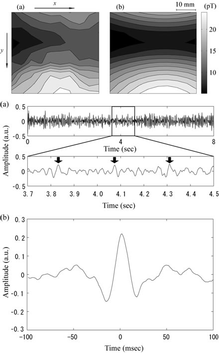
MEG (with superconducting quantum interference devices, SQUIDs) and high-field MRI (with superconducting magnets that require cryogenic cooling) are difficult to measure simultaneously. Optically pumped atomic magnetometers (OPAMs) are currently expected to overtake SQUIDs, and the possibilities for using OPAMs for biomagnetic field measurements and MRI have been demonstrated. We have developed a highly sensitive atomic magnetometer as a magnetic sensor to measure both MEG and MR signals. We describe the principles of the atomic magnetometer and the results of biomagnetic field measurements.
Optically pumped atomic magnetometers (OPAMs) using alkali metal vapors contained in glass cells are capable of measuring extremely small magnetic fields. OPAMS are based on the detection of Larmor spin precession in the alkali atoms contained in the glass cells. In recent years, OPAMs operating under spin-exchange relaxation-free (SERF) conditions have reached sensitivities comparable to and even surpassing those of SQUID-based magnetometers. The most sensitive atomic magnetometer has sensitivity in the subfemtotesla range (Kominis, Kornak, Allred, & Romalis, 2003). In addition, OPAMs have the intrinsic advantage of not requiring cryogenic cooling.