


- Non-invasive neuroimaging of higher brain functions
- Empirical research on brain mechanisms of visual perception and consciousness
- Development of an Ultra-Highly Sensitive Optically Pumped Atomic Magnetometer toward Non-invasive Brain Imaging
- Development of Integrative Multimodal Functional Neuroimaging Techniques
- Numerical methods for analyzing electric, magnetic, and electromagnetic fields in biological objects and their application to biological function engineering
Major research activities in our laboratory focus on the development of innovative methodologies for noninvasive imaging of the human brain.
Non-invasive neuroimaging of higher brain functions
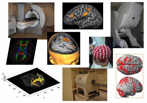
In addition to having an extensive knowledge of neuroscience, physics, chemistry, information science, and cognitive science, understanding the operation of the brain requires understanding and developing engineering techniques in measurement technology, imaging technology, and signal processing technology. Of these, non-invasive measurement techniques that do not damage the brain are particularly essential for investigating human brain functions.
In this lab, we are collaborating with Kyoto University's Graduate School of Medicine to make use of non-invasive measurement and imaging equipments, including 1.5 T and 3.0 T MRI systems, a 306-channel MEG system, as well as a high-resolution EEG system. Functional MRI: Neural activity in the brain is accompanied by relaxation of the arterioles, and an increase in the blood flow in the blood vessels in the activated parts of the brain results in a decrease in the deoxygenated hemoglobin concentration in the blood. This increases the transverse relaxation time of protons (hydrogen nuclei), which increases the intensity of a magnetic resonance signal. Such a signal is known as a blood oxygenation level dependent (BOLD) signal. fMRI is a technology for measuring brain functions by imaging BOLD signals. Although other (non-BOLD) fMRI devices have been trialed, most fMRI devices are still based on BOLD signals. MEG: When neural activity occurs, the electrical current flows inside the dendrite of the pyramidal cells in the cerebral cortex and generates very tiny magnetic field — approximately 100 million times weaker than the earth's magnetic field. In the 1960s, the invention of the SQUID — a supersensitive magnetic field detector utilizing the quantum interference effect inside a superconducting ring, such as a Josephson junction — for the first time made it possible to measure such tiny magnetic fields. A brain magnetic field measured using a SQUID is known as a magnetoencephalogram (MEG). From the magnetic field distribution measured in the vicinity of the scalp, by performing inverse analysis it is possible to estimate with high precision which parts of the brain are undergoing neural activity.